Monday, September 9, 2013

Teori Dasar X-Ray Diffraction (XRD)

Proses analisis menggunakan X-ray diffraction (XRD) merupakan salah satu metoda  karakterisasi material yang paling tua dan paling sering digunakan hingga sekarang. Teknik ini digunakan untuk mengidentifikasi fasa kristalin dalam material dengan cara menentukan parameter struktur kisi serta untuk mendapatkan ukuran partikel. Sinar X merupakan radiasi elektromagnetik yang memiliki energi tinggi sekitar 200 eV sampai 1 MeV. Sinar X dihasilkan oleh interaksi antara berkas elektron eksternal dengan elektron pada kulit atom. Spektrum sinar X memilki panjang gelombang 10-10  s/d 5-10 nm, berfrekuensi 1017-1020 Hz dan memiliki energi 103-106 eV. Panjang gelombang sinar X memiliki orde yang sama dengan jarak antar atom sehingga dapat digunakan sebagai sumber difraksi kristal. SinarX dihasilkan dari tumbukan elektron berkecepatan tinggi dengan logam sasaran. Olehk arena itu, suatu tabung sinar X harus mempunyai suatu sumber elektron, voltase tinggi, dan logam sasaran. Selanjutnya elektron elektron yang ditumbukan ini mengalami pengurangan kecepatan dengan cepat dan energinya diubah menjadi foton.
Sinar X ditemukan pertama kali oleh Wilhelm Conrad Rontgen pada tahun 1895, di Universitas Wurtzburg, Jerman. Karena asalnya tidak diketahui waktu itu maka disebut sinar X. Untuk penemuan ini Rontgen mendapat hadiah nobel pada tahun 1901, yang merupakan hadiah nobel pertama di bidang fisika. Sejak ditemukannya, sinar-X telah umum digunakan untuk tujuan pemeriksaan tidak merusak pada material maupun manusia. Disamping itu, sinar-X dapat juga digunakan untuk menghasilkan pola difraksi tertentu yang dapat digunakan dalam analisis kualitatif dan kuantitatif material. Pengujian dengan menggunakan sinar X disebut dengan pengujian XRD (X-Ray Diffraction).
XRD digunakan untuk analisis komposisi fasa atau senyawa pada material dan juga karakterisasi kristal. Prinsip dasar XRD adalah mendifraksi cahaya yang melalui celah kristal. Difraksi cahaya oleh kisi-kisi atau kristal ini dapat terjadi apabila difraksi tersebut berasal dari radius yang memiliki panjang gelombang yang setara dengan jarak antar atom, yaitu sekitar 1 Angstrom. Radiasi yang digunakan berupa radiasi sinar-X, elektron, dan neutron. Sinar-X merupakan foton dengan energi tinggi yang memiliki panjang gelombang berkisar antara 0.5 sampai 2.5 Angstrom. Ketika berkas sinar-X berinteraksi dengan suatu material, maka sebagian berkas akan diabsorbsi, ditransmisikan, dan sebagian lagi dihamburkan terdifraksi. Hamburan terdifraksi inilah yang dideteksi oleh XRD. Berkas sinar X yang dihamburkan tersebut ada yang saling menghilangkan karena fasanya berbeda dan ada juga yang saling menguatkan karena fasanya sama. Berkas sinar X yang saling menguatkan itulah yang disebut sebagai berkas difraksi. Hukum Bragg merumuskan tentang persyaratan yang harus dipenuhi agar berkas sinar X yang dihamburkan tersebut merupakan berkas difraksi. Ilustrasi difraksi sinar-X pada XRD dapat dilihat pada Gambar 1 dan Gambar 2.
Gambar 1 : Ilustrasi difraksi sinar-X pada XRD [1]

Gambar 2 : Ilustrasi difraksi sinar-X pada XRD [2]
Dari Gambar 2 dapat dideskripsikan sebagai berikut. Sinar datang yang menumbuk pada titik pada bidang pertama dan dihamburkan oleh atom P. Sinar datang yang kedua menumbuk bidang berikutnya dan dihamburkan oleh atom Q, sinar ini menempuh jarak SQ + QT bila dua sinar tersebut paralel dan satu fasa (saling menguatkan). Jarak tempuh ini merupakan kelipatan (n) panjang gelombang (λ), sehingga persamaan menjadi :


Persamaan diatas dikenal juga sebagai Bragg’s law, dimana, berdasarkan persamaan diatas, maka kita dapat mengetahui panjang gelombang sinar X (λ) dan sudut datang pada bidang kisi (θ), maka dengan ita kita akan dapat mengestimasi jarak antara dua bidang planar kristal (d001). Skema alat uji XRD dapat dilihat pada Gamnbar 3 dibawah ini.
Gambar 3: Skema alat uji XRD [3]
Dari metode difraksi kita dapat mengetahui secara langsung mengenai jarak rata-rata antar bidang atom. Kemudian kita juga dapat menentukan orientasi dari kristal tunggal. Secara langsung mendeteksi struktur kristal dari suatu material yang belum diketahui komposisinya. Kemudian secara tidak langsung mengukur ukuran, bentuk dan internal stres dari suatu kristal. Prinsip dari difraksi terjadi sebagai akibat dari pantulan elastis yang terjadi ketika sebuah sinar berinteraksi dengan sebuah target. Pantulan yang tidak terjadi kehilangan energi disebut pantulan elastis (elastic scatering). Ada dua karakteristik utama dari difraksi yaitu geometri dan intensitas. Geometri dari difraksi secara sederhana dijelaskan oleh Bragg’s Law (Lihat persamaan 2). Misalkan ada dua pantulan sinar α dan β. Secara matematis sinar β tertinggal dari sinar α sejauh SQ+QT yang sama dengan 2d sin θ secara geometris. Agar dua sinar ini dalam fasa yang sama maka jarak ini harus berupa kelipatan bilangan bulat dari panjang gelombang sinar λ. Maka didapatkanlah Hukum Bragg: 2d sin θ = nλSecara matematis, difraksi hanya terjadi ketika Hukum Bragg dipenuhi. Secara fisis jika kita mengetahui panjang gelombang dari sinar yang membentur kemudian kita bisa mengontrol sudut dari benturan maka kita bisa menentukan jarak antar atom (geometri dari latis). Persamaan ini adalah persamaan utama dalam difraksi. Secara praktis sebenarnya nilai n pada persamaan Bragg diatas nilainya 1. Sehingga cukup dengan persamaan 2d sin θ = λ . Dengan menghitung d dari rumus Bragg serta mengetahui nilai h, k, l dari masing-masing nilai d, dengan rumus-rumus yang telah ditentukan tiap-tiap bidang kristal kita bisa menentukan latis parameter (a, b dan c) sesuai dengan bentuk kristalnya.


Estimasi Crystallite Size dan Strain Menggunakan XRD

Elektron dan Neutron memiliki panjang gelombang yang sebanding dengan dimensi atomik sehingga radiasi sinar X dapat digunakan untuk menginvestigasi material kristalin. Teknik difraksi memanfaatkan radiasi yang terpantul dari berbagai sumber seperti atom dan kelompok atom dalam kristal. Ada beberapa macam difraksi yang dipakai dalam studi material yaitu: difraksi sinar X, difraksi neutron dan difraksi elektron. Namun yang sekarang umum dipakai adalah difraksi sinar X dan elektron. Metode yang sering digunakan untuk menganalisa struktur kristal adalah metode Scherrer. Ukuran kristallin ditentukan berdasarkan pelebaran puncak difraksi sinar X yang muncul. Metode ini sebenarnya memprediksi ukuran kristallin dalam material, bukan ukuran partikel. Jika satu partikel mengandung sejumlah kritallites yang kecil-kecil maka informasi yang diberikan metiode Schrerrer adalah ukuran kristallin tersebut, bukan ukuran partikel. Untuk partikel berukuran nanometer, biasanya satu partikel hanya mengandung satu kristallites. Dengan demikian, ukuran kristallinitas yang diprediksi dengan metode Schreer juga merupakan ukuran partikel. Berdasarkan metode ini, makin kecil ukuran kristallites maka makin lebar puncak difraksi yang dihasilkan, seperti diilustrasikan pada Gambar 4. Kristal yang berukuran besar dengan satu orientasi menghasilkan puncak difraksi yang mendekati sebuah garis vertikal. Kristallites yang sangat kecil menghasilkan puncak difraksi yang sangat lebar. Lebar puncak difraksi tersebut memberikan informasi tentang ukuran kristallites. Hubungan antara ukuran ksirtallites dengan lebar puncal difraksi sinar X dapat diproksimasi dengan persamaan Schrerer [5-9].
Gambar 4 : XRD Peaks [4]
Gambar 4 mengindikasikan bahwa makin lebar puncak difraksi sinar X maka semakin kecil ukuran kristallites. Ukuran kristallites yangmenghasilkan pola difraksi pada gambar bawah lebih kecil dari pada ukuran kristallites yang menghasilkan pola diffraksi atas. Puncak diffraksi dihasilkan oleh interferensi secara kontrukstif cahaya yang dipantulkan oleh bidang-bidang kristal. Hubungan antara ukuran ksirtallites dengan lebar puncal difraksi sinar X dapat diproksimasi dengan persamaan Schrerer [5-7].

Scherrer Formula

Dimana :
  • Crystallite size (satuan: nm) dinotasikan dengan symbol (D)
  •  FWHM (Line broadening at half the maximum intensity), Nilai yang dipakai adalah nilai FWHM setelah dikurangi oleh “the instrumental line broadening” (satuan: radian) dinotasikan dengan symbol (B)
  •  Bragg’s Angle dinotasikan dengan symbol (θ)
  • X-Ray wave length dinotasikan dengan symbol (λ)
  •  Adalah nilai konstantata “Shape Factor” (0.8-1) dinotasikan dengan symbol (K)
Perlu diingan disini adalah: Untuk memperoleh hasil estimasi ukuran kristal dengan lebih akurat maka, nilai FWHM harus dikoreksi oleh "Instrumental Line Broadening" berdasarkan persamaan berikut [4-9].
Dimana :
FWHMsample adalah lebar puncak difraksi puncak pada setengah maksimum dari sampel benda uji dan FWHMstandard adalah lebar puncak difraksi material standard yang sangat besar puncaknya berada di sekitar lokasi puncak sample yang akan kita hitung.
Contoh Estimasi Crystallite size menggunakan X-Ray Diffraction Analysis


Gambar 5: Penulis sedang melakukan sampel analisis menggunakan XRD Bruker 8 Advance 
Setelah data hasil uji sampel menggunakan XRD diperoleh, Data hasil analisa yang diperoleh tersimpan dalam format RAW.data, yang kemudian data tersebut dianalisa menggunakan Software EVA, data hasil uji sampel yang diperoleh adalah berupa peak seperti gambar dibawah ini.
Gambar 6: XRD Peak untuk sampel Fe powder  yang diuji penulis.


Sekilas Tentang Struktur Atom Suatu Unsur


Setiap atom terdiri dari inti yang sangat kecil yang terdiri dari proton dan neutron, dan di kelilingi oleh elektron yang bergerak. Elektron dan proton mempunyai muatan listrik yang besarnya 1,60 x 10⁻¹⁹ C dengan tanda negatif untuk elektron dan positif untuk proton sedangkan neutron tidak bermuatan listrik. Massa partikel-partikel subatom ini sangat kecil: proton dan neutron mempunyai massa kira-kira sama yaitu 1,67 x 10⁻²⁷ kg, dan lebih besar dari elektron yang massanya 9,11 x 10⁻³¹ kg. Setiap unsur kimia dibedakan oleh jumlah proton di dalam inti, atau nomor atom (Z). Untuk atom yang bermuatan listrik netral atau atom yang lengkap, nomor atom adalah sama dengan jumlah elektron. Nomor atom merupakan bilangan bulat dan mempunyai jangkauan dari 1 untuk hidrogen hingga 94 untuk plutonium yang merupakan nomor atom yang paling tinggi untuk unsur yang terbentuk secara alami. Massa atom (A) dari sebuah atom tertentu bisa dinyatakan sebagai jumlah massa proton dan neutron di dalam inti. Walaupun jumlah proton sama untuk semua atom pada sebuah unsur tertentu, namun jumlah neutron (N) bisa bervariasi. Karena itu atom dari sebuah unsur bisa mempunyai dua atau lebih massa atom yang disebut isotop. Berat atom berkaitan dengan berat rata-rata massa atom dari isotop yang terjadi secara alami. Satuan massa atom (sma) bisa digunakan untuk perhitungan berat atom. Suatu skala sudah ditentukan dimana 1 sma didefinisikan sebagai 1/12 massa atom dari isotop karbon yang paling umum, karbon 12 (12 C) (A = 12,00000). Dengan teori tersebut, massa proton dan neutron sedikit lebih besar dari satu, dan,
A Z + N
Berat atom dari unsur atau berat molekul dari senyawa bisa dijelaskan berdasarkan sma per atom (molekul) atau massa per mol material. Satu mol zat terdiri dari 6,023 x 1023 atom atau molekul (bilangan Avogadro). Kedua teori berat atom ini dikaitkan dengan persamaan berikut: 1 sma/atom (molekul) = 1 g/mol Sebagai contoh, berat atom besi adalah 55,85 sma/atom, atau 55,85 g/mol. Kadang-kadang penggunaan sma per atom atau molekul lebih disukai; pada kesempatan lain g/mol (atau kg/mol) juga digunakan.

Referensi :

  1. www.terrachem.de
  2. Callister,Jr, W.D., Rethwisch, D.G,. “Materials Science and Engineering An Introduction 8Th”, John Wiley & Sons, Inc. 2009.
  3. Saryanto, H., "High Temperature Oxidation Behavior of  Fe80Cr20 Alloys Implanted with Lanthanum and Titanium Dopant" Master Thesis, Universiti Tun Hussein Onn Malaysia, Malaysia, 2011.
  4. Abdullah, M & Khairurrijal,. "Review: Karakterisasi Nanomaterial" J. Nano Saintek. Vol. 2 No. 1, Feb. 2009.
  5. Abdullah, M., Isakndar, F., Okuyama, K. and Shi, F.G,. “ J. Appl. Phys. 89, 6431, 2001.
  6.  Abdullah, M. dan Khairurrijal, Nano Saintek. 1, 28. 2008.
  7.  Itoh, Y. Abdullah, M and Okuyama, K,.  J. Mater. Res. 19, 1077, 2004.
  8. P. Scherrer, “Bestimmung der Grösse und der inneren Struktur von Kolloidteilchen mittels Röntgenstrahlen,” Nachr. Ges. Wiss. Göttingen 26 (1918) pp 98-100.
  9. J.I. Langford and A.J.C. Wilson, “Scherrer after Sixty Years: A Survey and Some New Results in the Determination of Crystallite Size,” J. Appl. Cryst. 11 (1978) pp 102-113.

Failure Analysis


"In maintenance, when you get really good at something, you’re doing it much too often. There has to be a better way, and it’s time to do a serious failure analysis,"
In more than 90 percent of industrial cases a trained person can use the basic techniques of failure analysis to diagnose the mechanical causes behind a failure, without having to enlist outside sources and expensive analytical tools like electron microscopes. Then, knowing how a failure happened, the investigator can pursue the human roots of why it happened. There are times, however, when 90 percent accuracy is not good enough. When personal injury or a large loss is possible, a professional should guide the analysis.
To interpret a failure accurately, the analyst has to gather all pertinent facts and then decide what caused them. To be consistent, the analyst should develop and follow a logic path that ensures a critical feature will not be over looked. The following steps should be taken:
  1. Decide what to do. How detailed an analysis is necessary? Before starting, try to decide how important the analysis is. If the failure is relatively insignificant, in cost and inconvenience, it deserves a cursory analysis; the more detailed steps can be ignored. But this strategy increases the chance of error. Some failures deserve a 20-minute analysis with an 80 percent probability of being correct, but critical failures require true root cause failure analysis (RCFA), in which no questions are left unanswered. RCFA may require hundreds of man-hours, but it guarantees an accurate answer.
  2. Find out what happened. The most important step in solving a plant failure is to seek answers soon after it happened and talk to the people involved. Ask for their opinions, because they know the everyday occurrences at their worksite and their machinery better than anyone. Ask questions and try to get first person comments. Do not leave until you have a good understanding of exactly what happened and the sequence of events leading up to it.
  3. Make a preliminary investigation. At the site, examine the broken parts, looking for clues. Do not clean them yet because cleaning could wash away vital information. Document the conditions accurately and take photographs from a variety of angles of both the failed parts and the surroundings.
  4. Gather background data. What are the original design and the current operating conditions? While still at the site, determine the operating conditions; time, temperatures, amperage, voltage, load, humidity, pressure, lubricants, materials, operating procedures, shifts, corrosives, vibration, etc. Compare the difference between actual operating conditions and design conditions. Look at everything that could have an effect on machine operation.
  5. Determine what failed. After you leave the site and the immediate crush of the failure, look at the initial evidence and decide what failed first—the primary failure—and what secondary failures resulted from it. Sometimes these decisions are very difficult because of the size of analysis that is necessary.Find out what changed. Compare current operating conditions with those in the past. Has surrounding equipment been altered or revised? (Two failure examples on my desk have their mechanical roots in changes that took place years before the parts actually failed.)
  6. Examine and analyze the primary failure. Clean the component and look at it under low-power magnification, 5x to 50x. What does the failure face look like? From the failure face, determine the forces that were acting on the part. Were conditions consistent with the design? With actual operation? Are there other cracks or suspicious signs in the area of the failure? Important surfaces should be photographed and preserved for reference.
  7. Characterize the failed piece and the support material. Perform hardness test, dye penetrant and ultrasonic examination, lubricant analysis, alloy analysis, etc. Examine the failed part and the components around it to understand what they are. Check to see if the results agree with design conditions.
  8. Conduct detailed chemical and metallurgical analyses. Sophisticated chemical and metallurgical techniques may reveal clues to material weaknesses for minute quantities of chemical that may cause unusual fractures.
  9. Determine the failure type and the forces that caused it. Review all the steps listed. Leaving any questions unasked or unanswered reduces the accuracy of the analysis.
  10. Determine the root causes. Always ask, "Why did the failure happen in the first place?" this question usually leads to human factors and management systems. Typical root causes like "The shaft failed because of an engineering error" or "The valve failed because we decided not to PM it" or "The shaft failed because it was not aligned properly" expose areas where huge advances can be realized. However, these problems have to be dealt with differently; people will have to recognize personal errors and to change the way they think and act.

Definitions

  • Failure – when a person or component no longer performs as intended.
  • Primary Failure – the component that failed first and then caused secondary failures. Primary failures can usually be detected and monitored before they fail catastrophically. For example, a failure bearing may be the monitorable item that will, if neglected, eventually result in the secondary failure, the destruction of a gearbox.
  • Failure Investigation – an analysis of why something happened that does not delve as deeply into the causes as RCFA. As a result, the probability of an inaccurate diagnosis increases.
  • Fracture Face – the exposed surface where the failure actually progressed across the piece.Root Cause Failure Analysis – thorough analysis to find out why a failure occurred. It typically reaches into the human and management systems that allowed the failure to happen.
  •  Stress Concentrations – physical features that cause the apparent local stress in a part to be greater than the average across the piece. They can result from changes in shape, from defects, and from changes in metallurgy, and they can increase the local stress tenfold.

Types of failures
Different analysts use difference systems, but the most practical way for plant people to categorize failures is by overload, fatigue, corrosion-influenced fatigue, corrosion, and wear.
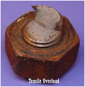
Overload:
·          Applying a single load causes the part to deform or fracture as the load is applied.

Figure 1: Close-Up overal view of bolt that failed by tensile overload/tensile bending fracture [1]

Fatigue: 
Fluctuating loads over a relatively long time causes this type of failure and usually leaves clues.
Figure 2
Results of high-temperature fatigue analysis of a prototype automotive piston show a failure as calculated by fe-safe. One design change to make the piston more fatigue resistant increased the radius of the fillet where the crack initiates Corrosion substantially reduces the fatigue strength of most metals and eventually causes failure at relatively light loads.

Apparently, the most common mode of failure in metallic parts is fatigue. Determination of a maintenance policy after the occurrence of a fatigue failure in service is based on finding the number of loading cycles that caused the failure [2]. The compression of gas within a plant is integral to the oil industry, too. It is thus of the utmost importance that premature failure of these pieces of equipment be avoided [3]. The rupture of metal parts is a complex phenomenon that depends on the nature of the material (composition, structure, and morphology), temperature, the deformation or excitation mode (traction, flexure, fatigue, etc.), and the rate at which strain is applied [4-5]. An examination of the fracture surfaces can provide information regarding the origin and cause of the fracture; these causes may include the apparent heterogeneity, ductility, and, sometimes, the grain size. When a metal piece breaks, two major questions should be answered: what are the modes and speed of rupture, and what is the origin of the damage (metallurgical failure, manufacturing defects, etc.) [4]. Damage tolerant fatigue design methods for critical rotating components, such as discs and shafts in gas turbine engines, are well established. These address crack propagation through the application of fracture mechanics, assuming defects are present in the material. The prediction methods have limitations; but at lower temperatures, these are conservative. It is not clear, however, if similar conservatism is valid at higher temperatures where additional failure modes due to creep and environment interact with those arising from fatigue [6].

Influenced fatigue:
Corrosion: 
The failure is the result of the electrical or biological action of the corrosion, causing a loss of material.
Wear: 
A variety of mechanisms result in loss of material by mechanical removal

Reference: 

  1. Jerner,R. C: Fastener Failure, http://www.metallurgist.com/html/FastenerFailures.htm
  2. Hershko, E., Mandelker, N., Gheorghiu, G., Sheinkopf, H., Cohen, I., and O. Levy, “Assessment of fatigue striation counting accuracy using high resolution scanning electron microscope,” Engineering Failure Analysis, 2008, pp. 20–27.
  3. Bennekom, A. van, Berndt, F., and Rassool, M.N., “Pump impeller failures—A compendium of case studies,” Engineering Failure Analysis, 2001, pp. 145–156.
  4. Boutarek, N., Saїdi, D., Acheheb, M.A., Iggui, M., Bouterfaїa, S., Competition between three damaging mechanisms in the fractured surface of an Inconel 713 superalloy,” Materials Characterization, 2007.
  5. Wei R.P. and Gangloff, R.P., Fracture Mechanics: Perspectives and Directions, Philadelphia: American Society for Testing and Materials, 1989.
  6. Hurley, P.J., Whittaker, M.T., Webster, P., and Evans, W.J., “A methodology for predicting creep/fatigue crack growth rates in Ti 6246,” International Journal of Fatigue, 2007, pp. 1702–1710.

Corrosion Theory

Corrosion Theory

Corrosion is the primary means by which metals deteriorate. Most metals corrode on contact with water (and moisture in the air), acids, bases, salts, oils, aggressive metal polishes, and other solid and liquid chemicals. Metals will also corrode when exposed to gaseous materials like acid vapors, formaldehyde gas, ammonia gas, and sulfur containing gases. Corrosion specifically refers to any process involving the deterioration or degradation of metal components.
When metal atoms are exposed to an environment containing water molecules they can give up electrons, becoming themselves positively charged ions, provided an electrical circuit can be completed. This effect can be concentrated locally to form a pit or, sometimes a crack, or it can extend across a wide area to produce general wastage. Localized corrosion that leads to pitting may provide sites for fatigue initiation and, additionally, corrosive agents like seawater may lead to greatly enhanced growth of the fatigue crack. Pitting corrosionalso occurs much faster in areas where microstructural changes have occurred due to welding operations.
The corrosion process (anodic reaction) of the metal dissolving as ions generates some electrons, as shown in the simple model on the left, that are consumed by a secondary process (cathodic reaction). These two processes have to balance their charges. The sites hosting these two processes can be located close to each other on the metal's surface, or far apart depending on the circumstances. This simple observation has a major impact in many aspects of corrosion prevention and control, for designing new corrosion monitoring techniques to avoiding the most insidious or localized forms of corrosion.

 Figure 1: Corrosion reaction


Corrosion sample
Shown below are typical examples of common corrosion failures
Fencing
This fence is located on a western-facing hillside a few miles from the coast.  As a result, it came in direct contact with the moist, salt-laden, onshore breezes.  The coating system applied to the fence was not robust enough for this environment.  The corroded areas were cut out and replaced.  The fence was stripped of its original coating and re-coated with a much thicker coating consisting of an epoxy primer followed by a urethane topcoat.
Figure 2: Fencing corrosion


Buried pipe
Two examples of water main failures are shown.
The first water main developed a leak after 8 years of service.  Although primed and tape-wrapped for corrosion protection, the pipe surface showed extensive pitting and corrosion damage.  In contrast, the inside of the pipe was undamaged.  The resistivity of the soil was measured.  Based upon the readings, the soil was found to be corrosive.  It was recommended that the water main be cathodically protected to prevent future
The second water main was in place since the 1920s.  It was bare cast iron, buried beneath the foundation of a home.  When the pipe began to leak it caused the surrounding soil to settle and the foundation of the home above it to crack.  In this instance, the hole in the water main started on the inside surface.

Figure 3: Buried pipe corrosion


Copper Water Lines
If the soil is corrosive enough it can even attack copper water lines as these examples show.  Typically, the surface of the water line at the bottom of the trench experiences the most corrosion.  The difference in oxygen concentration between the soil which was dug up to create the trench and then replaced (thus high oxygen content) and the undisturbed soil at the bottom of the trench (low oxygen content) creates a corrosion cell.  This cell, in addition to the soil’s general corrosiveness, causes the copper tube at the bottom of the trench to corrode.

Figure 4: Copper water line pipe corrosion

References :


5S (Bahasa Indonesia)

5S
5S adalah pondasi bagi TPM, oleh karenanya dasar keberhasilan penerapan TPM adalah berlangsungnya aktivitas 5S secara menyeluruh dan berkesinambungan. Bagi anda yang pernah berinteraksi dengan dunia industri tentunya tidak asing dengan istilah 5S. Perusahaan yang menerapkan program 5S akan terlihat bersih dan teratur.  Kondisi lingkungan kerja yang semrawut dan berantakan akan menyembunyikan masalah. Oleh karenanya program 5S dipandang sebagai usaha untuk memunculkan masalah yang selama ini tersembunyi  dari para pemecah masalah (problem solver). Saat ini, program 5S telah banyak diadopsi oleh berbagai industri di berbagai negara. Popularitas 5S ini tak lepas dari kesuksesan industri Jepang yang selama ini memusatkan  perhatiannya terhadap pengurangan segala  pemborosan (waste). 5S adalah landasan untuk membentuk perilaku manusia agar memiliki kebiasaan (habit) mengurangi pembororsan di tempat kerjanya.
Program 5S pertama kali diperkenalkan di Jepang sebagai suatu gerakan kebulatan tekad untuk mengadakan pemilahan (seiri), penataan (seiton), pembersihan (seiso), penjagaan kondisi yang mantap (seiketsu), dan penyadaran diri akan kebiasaan yang diperlukan untuk melaksanakan pekerjaan dengan baik (shitsuke). Masing-masing S dalam 5S beserta penjelasannya dijelaskan di bawah ini.

Tabel 1: Padanan 5S dalam Bahasa Indonesia dan Bahasa Inggris

1S-Seiri (Sorting): Ringkas
Seiri merupakan langkah awal implementasi 5S, yaitu: pemilahan barang yang berguna dan tidak berguna:
Simpan "Barang yang berguna"
Buang "Barang yang tidak berguna"
Dalam langkah awal ini dikenal istilah Red Tag Strategy,  yaitu menandai barang-barang yang sudah tidak berguna dengan label merah (red tag) agar mudah dibedakan dengan barang-barang yang masih berguna. Barang-barang dengan label merah kemudian disingkirkan dari tempat kerja. Semakin ramping (lean) tempat kerja dari barang-barang yang tidak dibutuhkan, maka akan semakin efisien tempat kerja tersebut.
       
      Contoh penerapan Seiri (Sorting): Ringkas

Figure 1: Sorting

2S-Seiton
Seiton adalah langkah kedua setelah pemilahan, yaitu: penataan barang yang berguna agara mudah dicari, dan aman, serta diberi indikasi.
Dalam langkah kedua ini dikenal istilah Signboard Strategy, yaitu menempatkan barang-barang berguna secara rapih dan teratur kemudian diberikan indikasi atau penjelasan tentang tempat, nama barang, dan berapa banyak barang tersebut agar pada saat akan digunakan barang tersebut mudah dan cepat diakses. Signboard strategy mengurangi pemborosan dalam bentuk gerakan mondar-mandir mencari barang.

3S-Seiso
Seiso adalah langkah ketiga setelah penataan, yaitu: pembersihan barang yang telah ditata dengan rapih agar tidak kotor, termasuk tempat kerja dan lingkungan serta mesin, baik mesin yang breakdown maupun dalam rangka program preventive maintenance (PM).
Sebisa mungkin tempat kerja dibuat bersih dan bersinar seperti ruang pameran agar lingkungan kerja sehat dan nyaman sehingga mencegah motivasi kerja yang turun akibat tempat kerja yang kotor dan berantakan.

4S-Seiketsu
Seiketsu adalah langkah selanjutnya setelah seiri, seiton, dan seiso, yaitu: penjagaan lingkungan kerja yang sudah rapi dan bersih menjadi suatu standar kerja. Keadaan yang telah dicapai dalam proses seiri, seiton, dan seiso harus distandarisasi. Standar-standar ini harus mudah dipahami, diimplementasikan ke seluruh anggota organisasi, dan diperiksa secara teratur dan berkala.

5S-Shitsuke
Shitsuke adalah langkah terakhir, yaitu penyadaran diri akan etika kerja:
                Disiplin terhadap standar
                Saling menghormati
                Malu melakukan pelanggaran
                Senang melakukan perbaikan
Suksesnya 5S terletak pada sejauhmana orang melakukan 5S sebagai suatu kebiasaan (habit) bukan paksaan sehingga inisiatif perbaikan akan muncul dengan sendirinya. Di bawah ini saya telah merangkum hal-hal penting untuk pelaksanaan program 5S berdasarkan beberapa literatur dan juga perspektif pribadi saat menyaksikan langsung aktivitas 5S di tempat kerja.
  • Membutuhkan keterlibatan/partisipasi semua orang dalam organisasi dari level atas sampai level bawah.
  • Membutuhkan komitmen manajemen untuk memastikan kegiatan 5S dilakukan setiap hari dan dianggap sebagai prioritas.
  • Merubah perspektif semua orang dalam organisasi bahwa 5S lebih dari sekedar program kebersihan maupun housekeeping management.
  • Menerapkan 5S secara konsisten untuk perubahan budaya.
  • Menggunakan sistem visual display untuk mengkomunikasikan  aktivitas 5S secara efektif.
  • Melakukan audit 5S secara teratur (mingguan, bulanan, dan surprise audit) untuk menilai performance.
  • Membutuhkan edukasi tentang konsep  dan keuntungan aktivitas 5S.
  • Sebagai penutup saya mau mengutip salah satu paragraf dari artikel yang disusun Utomo (2011) [1].
5S tidak sulit untuk dipahami, tapi 5S sangat sulit untuk dilaksanakan dengan benar. 5S memerlukan kegigihan, kebulatan tekad, dan memerlukan usaha yang terus menerus. 5S mungkin tidak akan memberikan hasil yang dramatis. Namun 5S membuat pekerjaan lebih mudah. 5S akan mengurangi pemborosan waktu kerja kita. 5S akan membuat kita bangga atas pekerjaan kita. 5S akan meningkatkan produktifitas kerja dan mutu yang lebih baik, sedikit demi sidikit, namun terus menerus.


Referensi :
  1. Utomo, A.C. (2011). Sejarah Singkat 5-S. Retrieved from http://www.scribd.com/doc/51971011/sejarah-singkat-5-S